Book an Appointment


Continuing the previous column on the causes and diagnosis of Cerebral Arteriovenous Malformation (AVM), this issue will share several real cases and targeted treatments.
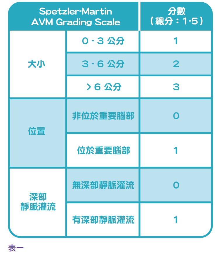
The factors in treatment decisions are based on the American Stroke Association’s standard of care (ASA Guideline), and we can use the research of American neurosurgeon Professor Spetzler-Martin to classify the risk of cerebral arteriovenous malformation hemangiomas into five levels (see Table 1) to distinguish treatment methods:
AVMs with low risk (first and second degree).
Neurosurgeons generally use minimally invasive microscopic excision, and if the location of malformed hemangiomas involves important cranial nerve functions, doctors may consider radiosurgery.
Moderate risk (Level III) AVM
Minimally invasive microscopic cerebrovascular surgical resection can be followed by endovascular embolization.
Highly risk (Level 4, 5) AVM
If the risk of any treatment option is high, the doctor will continue to observe conservatively.
Real case sharing
Case 1: Sudden rupture of malformed hemangiomas in women during pregnancy
Two girls, aged 15 and 16, who had always been healthy and had no headaches, discovered an aneurysm hidden in their brains in the early and second trimesters of pregnancy. Congenital cerebral arteriovenous malformation hemangioma suddenly rupture, resulting in severe bleeding, severe headache followed by rapid coma shock. After emergency brain surgery to remove blood stasis and reduce intracranial pressure, neurosurgeons waited three months to wait for the patient’s brain swelling to subside, and then treated with minimally invasive microscopic hemangioma resection surgery, and the two girls and their children made a complete recover.
Another 39-year-old woman in her late pregnancy was not so lucky. The night she was delivery a baby, her husband decisively rescued her and the baby is now a healthy and lovely child. Unfortunately, this woman, due to the rupture of arteriovenous malformed hemangioma, caused severe brain function damage, became a vegetative person, doctors tried various treatment options unsuccessfully, this lady was lying in the hospital for five years and finally died.
Case 2: Permanent damage to the brain of a four-year-old child
A four-year-old child, who has been healthy and normal since birth without any symptoms, suddenly felt severe headache while shopping with his mother one day, and then fell into a rapid coma. Sent to the hospital confirmed that it was a congenital brain arteriovenous malformation, and the hemangioma’s rupture caused severe hemorrhagic stroke. Although he was rescued, even after more than five years of treatment, he could not reverse the permanent damage to the brain caused by the stroke. Today, the child is still severely disabled, needs to stay in bed for a long time, and cannot communicate with others.
Case 3: Hemangioma: Rupture and bleeding of hemangioma, resulting in limb weakness
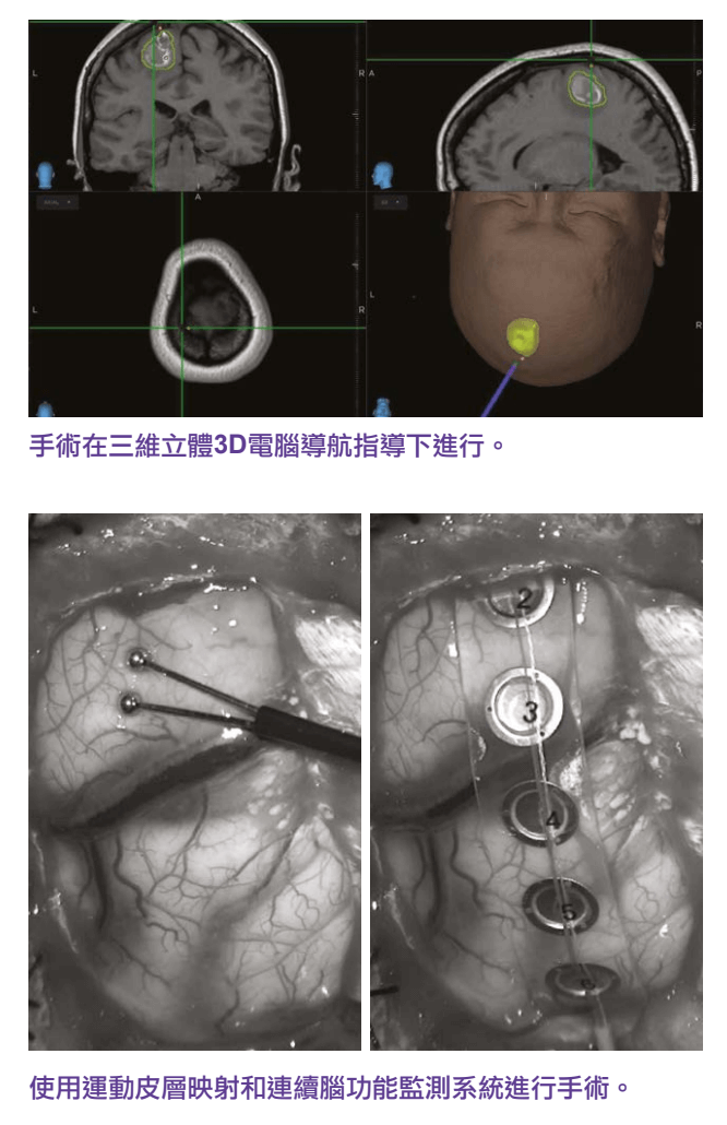
A 49-year-old woman felt weakness in her left leg two weeks after vaccination, and although she saw a Chinese medicine doctor and orthopedic surgeon, she had a MRI of the spine and could not find the cause. After a clinical evaluation, the neurosurgeon performed a 3D stereoscopic MRI cerebral angiography, which showed that her brain had congenital arteriovenous malformations and had been slightly bleeding, stasis the position of her right brain responsible for controlling the function of her left leg. Under the 3D stereoscopic computer navigation, motor neurocortical reflex and continuous brain function monitoring system, the neurosurgeon completely removed the hemangiomas and removed the blood clot under the microscope, and the nerve function of the patient’s brain was fully restored. After the surgery, the strength of the patient’s left leg returned to normal.
Case 4: Bleeding from malformed hemangioma causes severe cerebral seizures
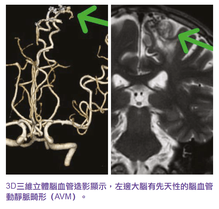
A 37-year-old woman was found fainting and unconscious at home and admitted to a public hospital. The patient and family were informed that the CT scan, EEG, and all blood tests were normal, and doctors still had no clinical clues to determine the cause of her condition. So after the patient was discharged, he consulted a neruosurgeon, and when the doctor reviewed the blood test report of the public hospital, he found that the patient’s serum muscle enzymes were very high, indicating that the cause of her fainting and unconsciousness was due to severe to brain epileptic seizures. The doctor then performed a 3D stereoscopic MRI cerebral angiogram, which showed that the patient’s left forehead brain lobe had a three-centimeter congenital arteriovenous malformation hemangioma and a small amount of recent bleeding, which was the cause of her loss of consciousness and epilepsy. Dynamic 3D Subtraction Cerebral Angiogram DSA determines blood flow patterns within hemangiomas. After four hours of microscopic surgery, the vascular surgeon saw the old residual blood of hemosiderin in the brain next to the hemangioma, and the hemangioma’s and bruising were completely removed. The patient’s brain nerve function was well preserved, and he was discharged from the hospital two days after the operation, and there were no seizures of epilepsy in 10 years.
Case 5: Premature occlusion of aneurysm by radiotherapy led to aneurysm rupturing
When a 42-year-old housewife went to Shenzhen with her family for a full body examination, she found a congenital cerebral arteriovenous malformation aneurysm in her brain. Upon her return to Hong Kong, the woman sought medical treatment in a public hospital and was told by the doctor that she should adopt an observation and conservative treatment plan. However, under the persuasion of his family, the patient went to Guangzhou for X-ray knife radiotherapy. Shortly after returning to Hong Kong after surgery, the patient who was sleeping and suddenly get into the convulsion stage in the morning and was sent to a nearby public hospital for emergency treatment, after brain surgery craniotomy to remove blood clot and reduce intracranial pressure, neurosurgeons conducted a detailed investigation, suspected that the position of radiotherapy was not accurate enough, resulting in premature occlusion of the veins of the malformed blood vessels, so that blood would only flow into the malformed blood vessels through the arterial vessels, and could not flow out through the occluded venous vessels, which increased the blood pressure in the malformed aneurysm to burst, resulting in hemorrhagic stroke. Although the patient was fortunate enough to be saved, the damaged brain nerve area caused her to lose the ability to speak (commonly known as “aphasia") and the mobility of her right hand.
Case 6:
The patients are a 16-year-old boy and a 56-year-old man, both of whom underwent a thorough examination of the brain and cerebrovascular structures without any symptoms. 3D three-dimensional MRI cerebral angiography showed that both brains had congenital cerebrovascular arteriovenous malformations aneurysm, under the advice of neurosurgeons, after a period of observation and conservative treatment, and then the use of minimally invasive cerebrovascular catheter occlusion surgery and radiation therapy, the aneurysm were effectively cured, and the patient’s innate brain timing bomb was removed, eliminating their risk of hemorrhagic stroke in the future.
The sharing and treatment content of this article is for general and reference purposes only, with the aim of providing patients and their families with a certain understanding of the disease and different treatment options before deciding to undergo treatment. Patients need to make appropriate treatment decisions based on their actual situation, and this overview does not provide individual patients with a definitive diagnosis or recommended treatment options. Any treatment plan has its potential risks, and patients are requested to fully discuss and cooperate with their healthcare professionals during their visit.