Book an Appointment


2013-04-15
Case Sharing
55-year-old Ms. Ko, a full-time housewife, had low back pain lasting for four months, and also developed symptoms of numbness of her left foot and toes.
Ms. Ko did not get better after four weeks of physical therapy. The orthopaedic surgeon took the patient’s MRI and found that the lumbar spine was severely degenerated from the second to the fifth.
In addition, a 20x12x13mm synovial cyst was found on the small joint between the fourth and fifth lumbar spine. The cyst also blocked the position of the left spinal canal and pressed the left nerve root.
Ms. Ko received COX® decompression manipulation. After 18 COX® treatments, her back pain and foot pain/numbness disappeared. Now the decompression therapy is maintained every three months, and the situation has settled down.
Case Analysis
Facet joint synovial cyst is a small fluid-filled cyst, which is generally a benign cyst. It is usually found in patients over 45 years of age or older, especially in people 65 years of age or older.
This synovial cyst is caused by severe degeneration of the spine. Due to joint degeneration, the surrounding synovial fluid increases, and the increased synovial fluid enters the synovial membrane of the joint to form a synovial sac. The synovial sac contains inflammatory cells, fibrin and hyperplasia.
Clinical symptoms of Lumbar facet joint synovial cyst
The patient’s symptoms are very similar to those of a herniated disc, with claudication such as sciatica or spinal stenosis.The patient’s low back pain and foot pain also change according to the patient’s posture. When the patient sits down, the pain will ease a little, but when he walks again or straightens the waist, the waist and feet pain reappears.The reason is that when the patient sits down, the space where the cyst presses on the spinal canal opens, and the compressed nerve root can reduce the pressure, so the pain will be relieved.
Diagnose of Lumbar facet joint synovial cyst
MRI scanning can clearly show the location, distribution and size of synovial cysts. X-rays are difficult to see the image of the cyst.

The location of the synovial cyst in the facet joint (magnetic resonance imaging in straight view)
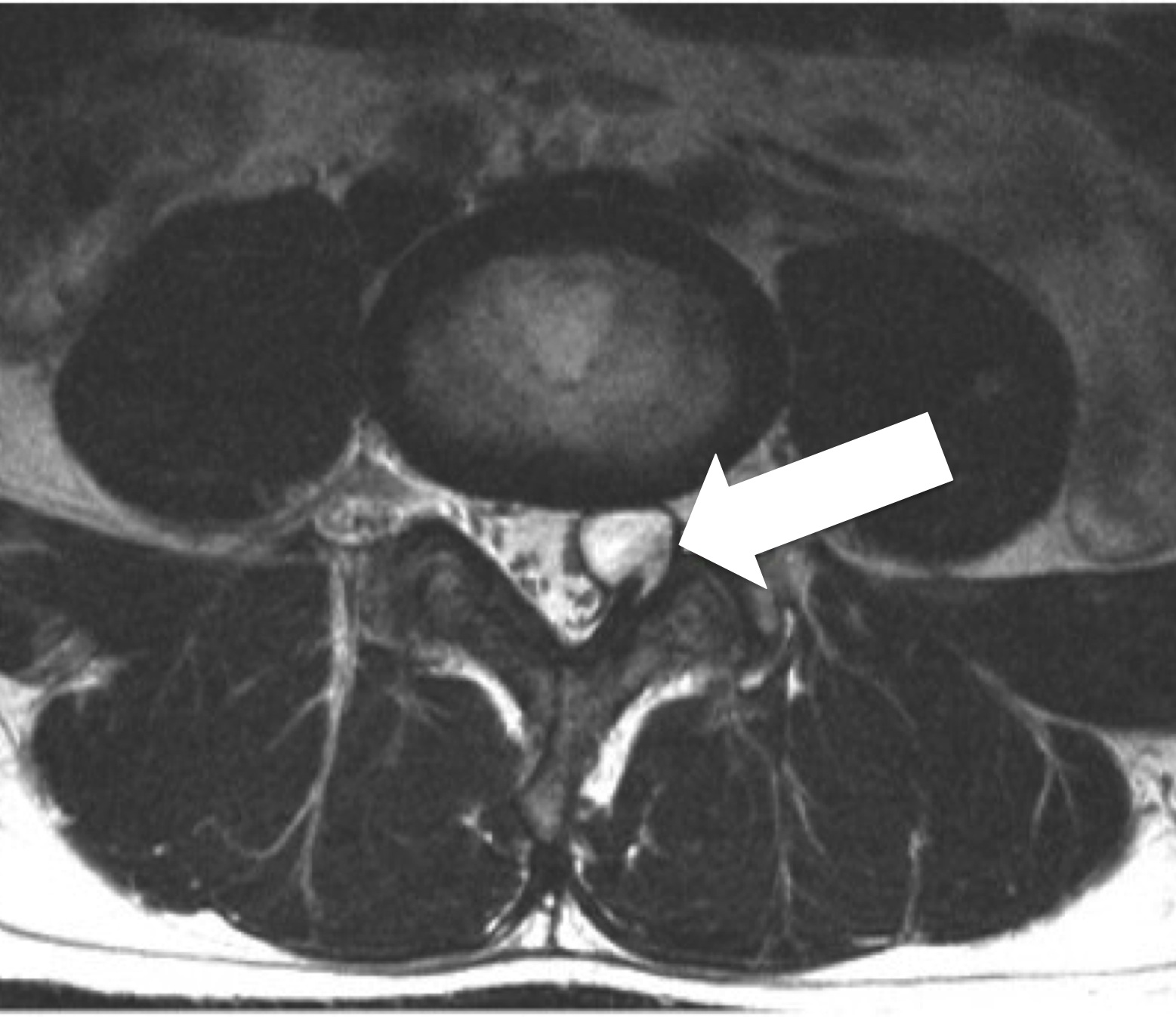
Location of facet joint synovial cyst (cross-sectional magnetic resonance image)
Treatment
Continued treatment: COX® decompression manipulation can open the spinal canal to reduce nerve root pressure, increase blood circulation in the affected area and relieve the patient’s low back pain and foot numbness. Generally, after about 12 treatments, the patient’s low back pain and foot pain should be reduced by 30-50%. If the patient has no significant progress, the patient will be referred to neurosurgeon for surgery option.
Neurosurgical treatment: Minimally invasive spinal decompression surgery is used to reduce nerve root compression. Non-surgical methods include injection of steroids in the lumbar facet joints or puncture of cysts.