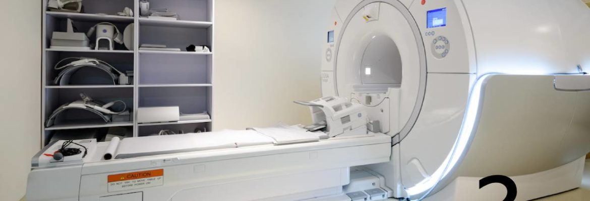
Magnetic resonance imaging show herinated disc
Why do doctors recommend an MRI scan instead of an X-ray when the doctor thinks that the patient may have a herniated disc? Let us talk about the reasons.
When dealing with a herniated disc, X-rays can only capture bone lesions of the spine, including structure, alignment, subluxation, degeneration and bone spurs, etc. The MRI scan can see the structure and water contents in the intervertebral disc, the condition of herniated or prolapsed intervertebral disc, the severity of nerve compression and can also see the condition of spinal canal stenosis due to spinal degeneration. Therefore, to diagnose a herniated disc, a MRI scan is necessary.
Case 1: A 39-year-old professional model
39-year-old David is a professional model. In February of this year, he sprained his neck while doing weightlifting. At first he thought it was a muscle inflammation, but two weeks later he still felt soreness and numbness in his neck and hands. After receiving an x-ray examination, there were no bone lesions or narrowing of the vertebral foramen on the x-rays. It did not seem to be a symptom of a herniated disc. The doctor prescribed anti-inflammatory, pain-relieving and muscle-relaxing drugs. After starting to take the drug, it relieved a bit, but after two months, he has atrophy of the upper arm and right shoulder muscles, and the upper arm was unable to lift weights. He felt body began to feel weak! After I took his medical history and perform physical examination on him, I believed the diagnosis is cause by the herniated disc. As in the X-ray the C6,7 were slightly narrower, and the X-ray of the foramen was not narrowing. I believe that X-ray alone can not diagnose his condition. Therefore I did an MRI scan in clinic and found out his C6,7 has a large herniated disc pressing against the right nerve root. This is the cause of his right shoulder, upper back, hand muscle atrophy and right hand weakness. There is also a herniated disc between C5,6 which is also causing him to have numbness in his right hand.
X-ray only show C6,7 vertebrae space is narrower
C6,7 has a large herniated disc pressing against the right nerve root. There is also a herniated disc between C5,6 which is also causing him to have numbness in his right hand.
Case 2: 71-year-old retire lady
71-year-old Ms. Cheung is a retired person who loves mountain climbing and photography. In recent months, she has found pain and numbness in her left foot. The symptoms are weakness and numbness in her left foot after walking for a long time, and a dull pain in the left pelvic bone. From the X-ray taken by the doctor, can see the lumbar scoliosis, the pelvis is slightly higher, the entire lumbar spine is degenerated and it is diagnosed she has L5,S1 disc herniation.
I did an MRI scan in the clinic, and found that her symptoms were not due to herniated disc L5,S1 level (although the space between L5,S1 was really narrowing from the x-ray view). Instead, her herniated disc is located at L3,4 and there is spinal stenosis observed at this level, the L3,4 herniated disc pressed on the left nerve root, causing her left foot to be numb and painful.
X ray only show pelvic tilt and L5,S1 disc space narrowing.
MRI actually show L3,4 disc herniation (red arrow), no L5,S1 disc herniation (yellow arrow) that was suspected in X ray
MRI scan can diagnose whether the intervertebral disc is herniated, and understand the condition of the spinal nerves
MRI can be used to diagnose disc disease
Based on the sharing of the above two cases, we can see from the MRI scan that we can clearly distinguish which intervertebral disc herniation, which spine has spinal stenosis and which spinal nerve is compressed! If you only look at the x-ray to observe the curvature of the spine and the distance of the intervertebral disc , spondylolisthesis, bone spurs and narrowing of the vertebral foramen, or doing designated muscle and nerve reflex tests, can not confirm whether the patient has the symptoms of a herniated disc.
MRI scans must be used to diagnose whether the patient has herniated disc and understand the spinal nerves condition in order to provide the best treatment to patients.

 Book an Appointment


