Book an Appointment


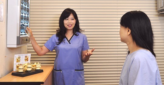
Sciatica caused by disc protrusion ( part II)
Patients with disc protrusion and herniated disc, the pain is usually located in the buttocks and legs (see the previous issue for symptoms). Because the pain and numbness are very severe, the first reaction of the patient is to take painkillers and anti-inflammatory medication to relieve the pain, but after medication effectiveness subside, the pain usually still persist because the disc is still pressed on the nerve root. Of course, if the patient’s pain is really unbearable, painkillers are still needed.
Most of them have done physical therapy, acupuncture, massage, injection and medicine, and are even considering or have undergone discectomy surgery (one or more times). Patients often come with a large stack of X-ray films and MRI scanning. In fact, the treatment of disc protrusion takes a long time. Patients need great patience and endurance to do the long treatment. After recovery, many lifestyle habits must be changed. To effectively treat disc protrusion, the internal pressure of disc must first be reduced.
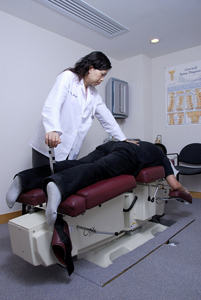
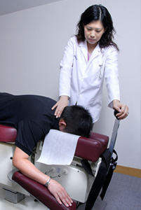
COX® Decompression Manipulation
COX® Decompression Manipulation is a set of treatments invented by the American chiropractor Dr. James M. Cox in the 1960s and has been used for more than 40 years. In academic research, a number of verifications have also been made to support its effective treatment effect (* Please refer to a number of scientific research studies of COX® Technic). COX® Decompression Manipulation uses a special decompression bed, and then presses the patient’s disc protruding section for decompression. After decompression, the soft nucleus pressure reduced. The space for the spinal foramen increased, and at the same time the compressed nerve roots get released, so the patient’s leg pain or leg numbness can be improved.
After many months of treatment, the pressure in the cartilage will continue to decrease, allowing enough time for the fibrous annulus in the outer layer of cartilage to repair. However, if the annulus fibrosus is not completely repaired, when the cartilage is compressed again, the annulus fibrosus will be torn again, the nucleus pulposus will continue to overflow, and the nerve root will be pressed to produce pain again. Therefore, patients cannot carry heavy objects, do strenuous exercises, stand for long periods of time, sit for a long time, and cannot do fitness, yoga, ball games, dancing or swimming. The patient can only try to rest appropriately until entering the rehabilitation stage (clinical experience is about 3 months).
When the patient’s sciatica symptoms have completely disappeared, MRI is often required to verify whether the cartilage can be completely reset, and to see if the cartilage is protruding again. I have repeatedly emphasized this point. Cartilage will degenerate and become dehydrated with age. Although the cartilage protrusion remains the same, the important point is that the soft bone marrow nucleus has no longer prolapsed or overflowed to compress the nerve roots, so the patient’s pain disappeared.
After recovery, patients are most worried about the recurrence of cartilage protrusion. Although the condition is controlled, if the patient don’t have correct posture, doesn’t do strengthen exercise to exercise muscles, nor improve their living habits, the cartilage protrusion will most likely occur again.